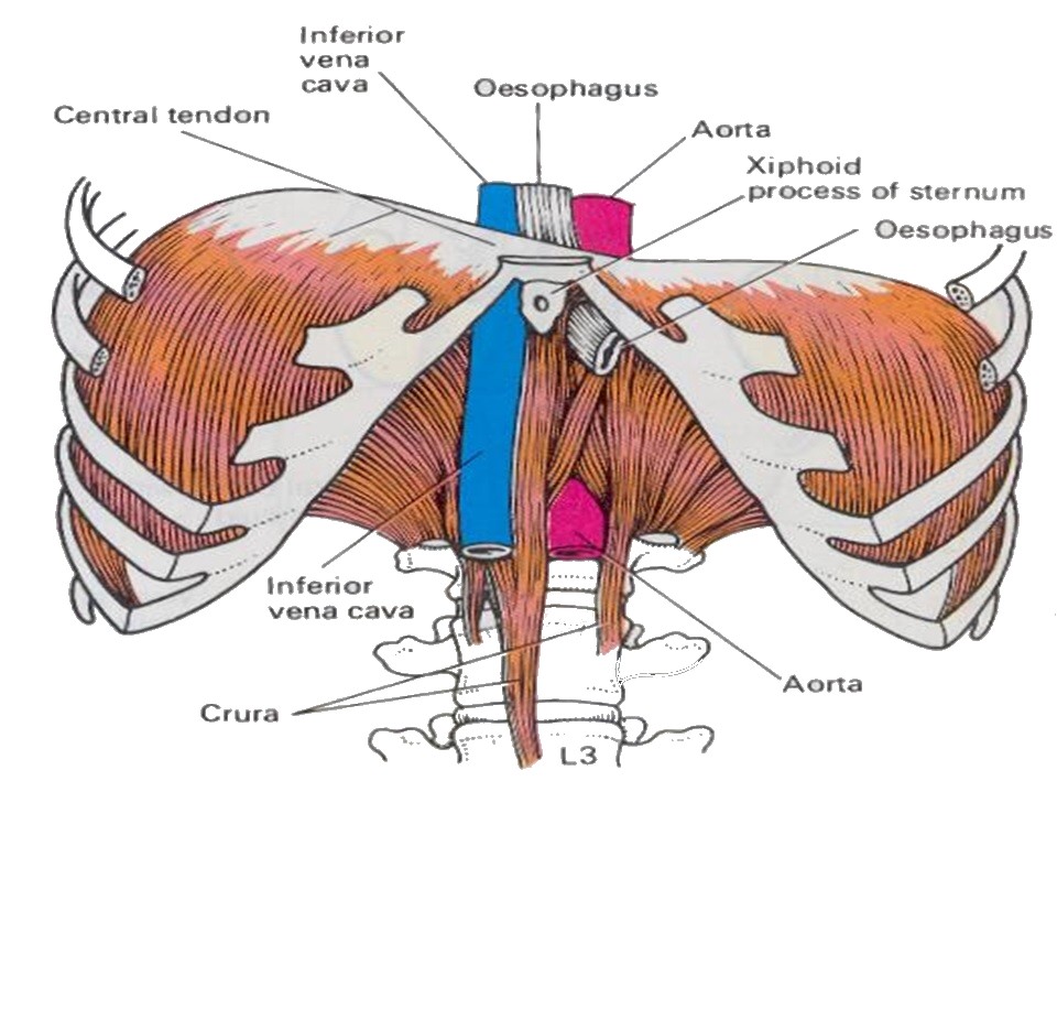
Diaphragm
Definition
Dome shaped structure separating the thoracic and abdominal cavities.
It is the floor of the thoracic cavity and the roof of the abdominal cavity
Features
Has a central tendon
From the central tendon muscle fibres arise
Get attached to the lower ribs and sternum.
To the vertebrae it is attached by two crurae.
When the diaphragm is relaxed the central tendon is at the level of the T8 vertebra.
When it contracts its muscle fibres shorten and the central tendon is pulled downwards, enlarging the thoracic cavity in length.
Openings
Three large openings
the aortic
the esophageal
the caval opening
a series of smaller ones
Aortic hiatus : T12 - The aorta does not pierce the diaphragm but rather passes behind it in between the left and right crus.
Is in the posterior part of the diaphragm, between the left and right crus.
It contains the aorta, the azygos vein, and the thoracic duct
Esophageal hiatus : contains the esophagus, and anterior and posterior vagal trunks.
.Caval Opening : The inferior vena cava passes through this opening
two lesser apertures of right crus : greater and lesser right splanchnic nerves
two lesser apertures of left crus : greater and lesser left splanchnic nerves and the hemiazygos vein
behind the diaphragm, under the medial lumbocostal arch : sympathetic trunk
Nerve supply
Phrenic nerve C4